Why? (The Short Story)
Can optics achieve the next frontier in healthcare? Radiation-free and non-invasive diagnostics? The motivation of this research focus is to facilitate low-cost diagnostics and extend the understanding of tissue-light interactions across in vivo and synthetic tissue models.
The Long(er) Story
Biophotonics and Diagnostics
Optical approaches could allow future medical diagnostics to be capable of assessing conditions within the human body in non-invasive and/or non-contact approaches. Our research in this domain is focused on devising non-contact approaches to diagnostics, understanding vital signs without invasive approaches and understanding the anatomical impacts of permanent medical conditions.
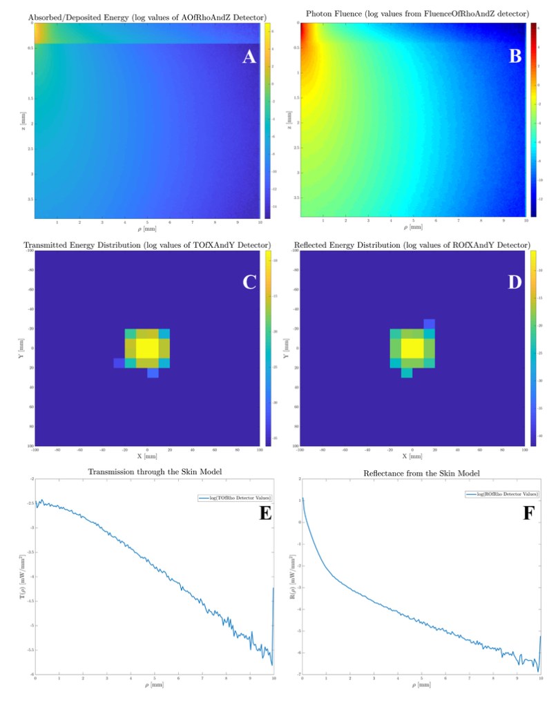
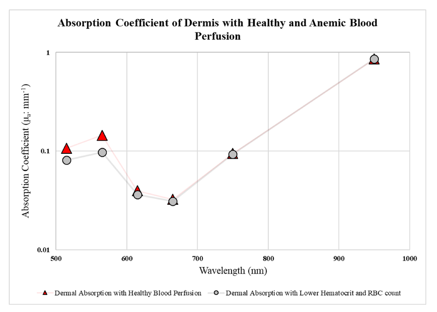
Our current research builds on identifying markers for anaemia (SPIE 2020 Publication and Dataset), application of Monte Carlo methods and current diagnostic techniques such as ultrasound imaging for more accurate models (Journal of Biophotonics, Dataset) and device fabrication for blood perfusion monitoring in skin flaps and hypertension.
Ultrasound-based Diagnostics
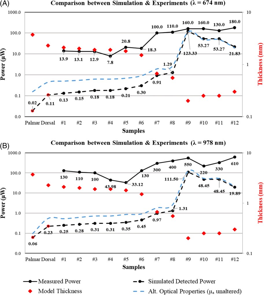
Diagnosis of skin cancer is currently done using direct observation, biopsies and histology analysis of the excised tissue. Subsequently, the affected tissue is treated with either invasive surgical procedures or non-invasive photodynamic therapy. These procedures are expensive and could benefit from a more targeted approach. Our research, published in the Journal of Biophotonics (2022), presents a novel approach of using high-resolution, high-frequency ultrasound imaging to accurately determine the geometry of the tumours and build optical models to plan strategies with the least exposure to PDT and accurate surgical and/or light-based interventions.
Human Skin Equivalents
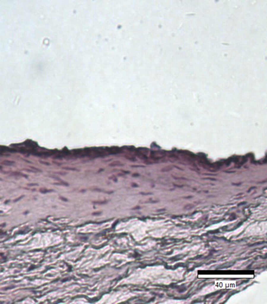
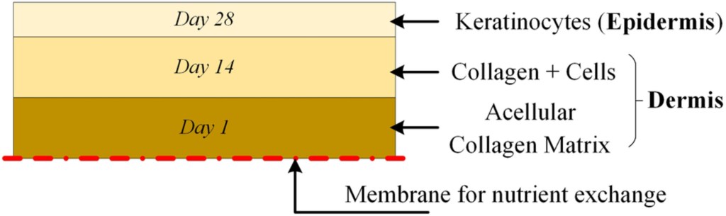
Human skin equivalents (HSEs) are live, three-dimensional skin models cultured in the lab to replicate the biological interactions and behaviour of skin in vivo. These models allow a varied set of applications from drug response monitoring to plastic surgery and act as an alternative to animal models. For these models to be used extensively, their behaviour must be similar to human skin, optically and mechanically. Our investigations into HSEs has led to a pilot study, bridging two domains of research — biophotonics and tissue engineering. In collaboration with Dr Sarah Junaid (Aston University), we presented a novel study for assessing optical transport through tissue, as a comparison with in vivo skin. Our research, published in the Journal of Biophotonics, has been covered by the Institute of Physics in Engineering and Medicine’s SCOPE and IEEE Photonics Society’s Newsletter.
“Biophotonics for all” Outreach Kit
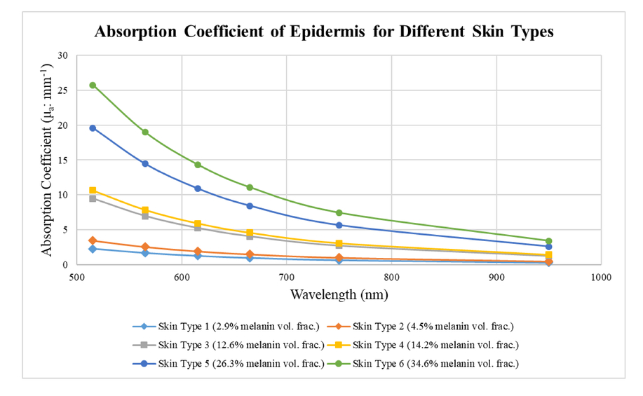
Biophotonics is becoming increasingly prominent in value and visibility. What was once an advanced field of research is now being included increasingly in undergraduate and postgraduate programmes. To increase biophotonics-related STEM awareness, we developed simple and inexpensive LEDs and a camera set-up that allows visualising the blood vessels beneath the skin. The kit uses inexpensive blue, green, red and near-infrared LEDs to show the absorption of shorter wavelengths and transmission in the longer wavelengths in the finger. As an outreach and educational tool, this kit will illustrate the potential of using light for diagnostics.
This kit will be presented at the 2023 Education and Training in Optics and Photonics (ETOP) conference, and available subsequently through SPIE proceedings.
Relevant Publications and Datasets
Gibson G. and Kallepalli A., Biophotonics For All: Light transport through tissue, Education and Training in Optics and Photonics (ETOP) 2023 (Cocoa Beach, Florida); 16 May 2023
M. Main and A. Kallepalli, Towards point-of-care diagnostics and monitoring of hypertensive episodes (A Monte Carlo approach), in Biophotonics Congress: Optics in the Life Sciences 2023 (OMA, NTM, BODA, OMP, BRAIN), Technical Digest Series (Optica Publishing Group, 2023)
Kallepalli A. et al., An ultrasonography-based approach for tissue modelling to inform phototherapy treatment strategies., J. Biophotonics, 2022; DOI: 10.1002/jbio.202100275
Preprint accessible on arXiv
Kallepalli A. et al., Optical investigation of three‐dimensional human skin equivalents: A pilot study. J. Biophotonics. 2020; 13:e20190053; DOI: 10.1002/jbio.201960053
Kallepalli A. and James D. B., Quantification and influence of skin chromophores for remote detection of anaemic conditions. Proc. SPIE 11238, Optical Interactions with Tissue and Cells XXXI, 112381B (20 February 2020); DOI: 10.1117/12.2545784
Kallepalli A., and Gibson G. (2023); Biophotonics for All: Light transport through tissue. Zenodo. DOI: 10.5281/zenodo.7876304
Outreach Tools and Kits
Kallepalli A., James D. B., Richardson M. A. (2021): Monte Carlo simulation results for full finger models based on ultrasound image data. Cranfield Online Research Data (CORD). DOI: 10.17862/cranfield.rd.11603883.v2
Kallepalli A. and James D. B. (2020): Monte Carlo simulation results for anaemia detection in the skin. Cranfield Online Research Data (CORD). DOI: 10.17862/cranfield.rd.11317187.v2